Español
Phenotyping the central nervous system of the embryonic mouse by magnetic resonance microscopy
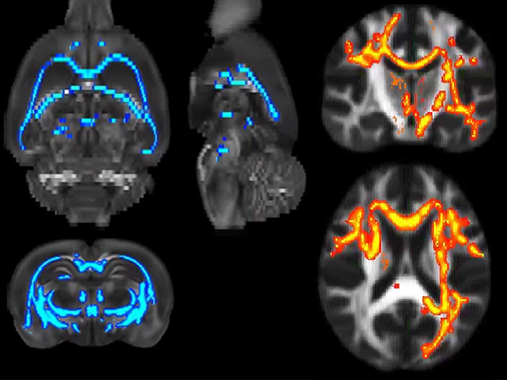
Genetic mouse models of neurodevelopmental disorders are being massively generated, but technologies for their high-throughput phenotyping are missing. The potential of highresolution magnetic resonance imaging (MRI) for structural phenotyping has been demonstrated before. However, application to the embryonic mouse central nervous system has been limited by the insufficient anatomical detail. Here we present a method that combines staining of live embryos with a contrast agent together with MR microscopy after fixation, to provide unprecedented anatomical detail at relevant embryonic stages. By using this method we have phenotyped the embryonic forebrain of Robo1/2-/- double mutant mice enabling us to identify most of the well-known anatomical defects in these mutants, as well as novel more subtle alterations. We thus demonstrate the potential of this methodology for a fast and reliable screening of subtle structural abnormalities in the developing mouse brain, as those associated to defects in disease-susceptibility genes of neurologic and psychiatric relevance.