
 Español
Magnetic Resonance and Molecular Imaging
The Magnetic Resonance and Molecular Imaging Facility provides state-of-the-art Magnetic Resonance (MR) equipment and scientific advice in MR to public and private research institutions.
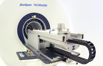
The facility was created in 2011 as a central service in the Instituto de Neurociencias (CSIC/UMH). The service has a Bruker BioSpec 7Tesla (30 cm internal diameter) fully equipped to perform in vivo and ex vivo MR Imaging and Spectroscopy. The service is equipped with volume coils for rodent whole body imaging and single voxel spectroscopy. Also it has a special set up for brain imaging using a phase array coil, optimized for functional Magnetic Resonance Imaging (fMRI).
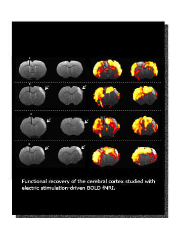
The service provides necessary instrumentation to anesthetize the animals using inhalation or injectable anesthesia. Equipment for non-invasive and fully MR-compatible physiology monitoring during imaging acquisition is also available, including body temperature, arterial pressure, heart and breath rate and oxygen saturation. A 4 channel electric stimulation device for stimulation-driven fMRI is available. Additional equipment to perform surgery and artificial ventilation could be provided upon request.
Other activities
The service organizes and participates in events related to formation and divulgation of the technique. For that, general talks are organized periodically about security, basics of magnetic resonance theory and applications. Personalized courses could be also arranged under specific request.
Also, it’s integrated in the general activities organized by the Neuroscience Institute including visits of students, week of science, Brain Awareness Week, open doors journeys, seminars, etc.
Photo Gallery
The service has a Bruker BioSpec 7Tesla (30 cm internal diameter) fully equipped to perform in vivo and ex vivo MR Imaging and Spectroscopy. The service is equipped with volume coils for rodent whole body imaging and single voxel spectroscopy. Also it has a special set up for brain imaging using a phase array coil, optimized for functional Magnetic Resonance Imaging (fMRI).
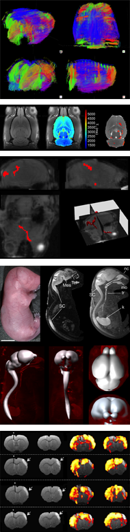
Imaging modalities performed by Molecular Imaging facility:
- Functional Magnetic Resonance Imaging (fMRI)
- Anatomic Spin Eco (SE) and Gradient Eco (GE) images weighted in T1, T2 and spin density images
- Diffusion weighted images (DW), apparent diffusion coefficient map (ADC) and diffusion tensor imaging (DTI)
- Spin Echo and Gradient Eco 3D images
- Angiography
- In vivo localized spectroscopy
- Perfusion weighted brain imaging
Applications
Neuroimaging
- Brain activity
- Morphometry
- Angiography
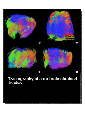
- Tractography
- In vivo brain metabolism
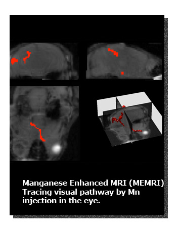
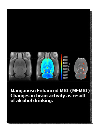
- MEMRI (manganese enhanced MRI)
- Evaluation of animal models of neurodegenerative diseases
- Evaluation of animal model of cerebral ischemia
- Neurotoxicity and neuroprotection
Transgenic animal characterization
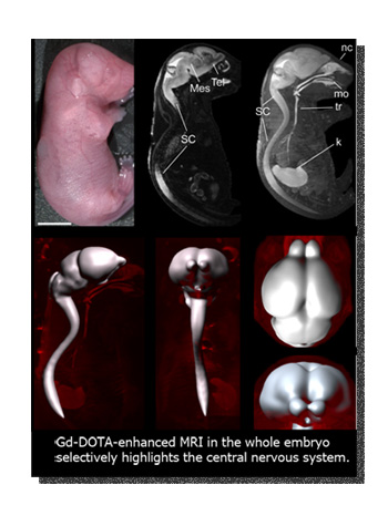
- Post partum morphometry
- Brain activity (fMRI)
- In vivo metabolism
- Tractography (DTI)
- Multiparametric imaging
Experimental oncology
- Morphology of oncogenesis in experimental models
- Angiogenesis: blood flow and vascular volume
- Capillary permeability
- Comprehensive assessment of tumor models
- Multiparametric imaging
List of Services Offered
MR fares have been calculated according to the different items to be defrayed by different types of user:
- Internal users. Service maintenance and consumables will be defrayed. Staff Researchers from CSIC and UMH will be considered as internal users.
- Users from other Public Research Institutions (OPIs). Rates are calculated according to costs of Service maintenance, consumables and the estimated cost of equipment repairs.
- Companies and private institutions. They will be charged according to market prices, and in any case, rates will be estimated to real costs (including expenses of staff involved in the studies).
Table 1. Price list
| Type of user | Items to cover | €/h (working hours) |
Overnight | Weekends |
| Internal (CSIC & UMH) |
Maintenance and consumables |
31 | 100 | 150 |
| OPIs | Maintenance, consumables and repairs |
65 | 200 | 250 |
| Companies and private institutions |
Maintenance, consumables, repairs and staff |
150 | 300 | 400 |
Animal studies will be charge with additional 10€/h in anesthesia.
For those studies that require long time acquisition and no requirement for continuous supervision (more than 10h, as with phantoms and other non-living samples), special prices will be applied, as depicted in the Price list (table 1, ‘Overnight’ and ‘Weekends’).
If image processing or analysis is required, the same rates (€/h) will be applied depending on the time consumed in the process.
The unit will also provide technical and scientific consulting, as well as software development for special applications, upon demand.
Scientific Director:
Dra. Silvia De Santis
dsilvia@umh.es
Technical Manager:
Dr. Mohamed Kotb Selim
mselim@umh.es
Tel: 965 91 9223
Technician:
Víctor Sanchís Sala
v.sanchis@umh.es
Tel: 965 91 9223
Magnetic Resonance and Molecular Imaging Service
Instituto de Neurociencias
Universidad Miguel Hernández – CSIC
Campus de San Juan
Sant Joan d’Alacant
Alicante | España
Tel. + 34 965 23 37 00
(ext. service 9223)