Español
Microstructural White Matter Alterations in Men With Alcohol Use Disorder and Rats With Excessive Alcohol Consumption During Early Abstinence
Importance:
Although the detrimental effects of alcohol on the brain are widely acknowledged, observed structural changes are highly heterogeneous, and diagnostic markers for characterizing alcohol-induced brain damage, especially in early abstinence, are lacking. This heterogeneity, likely contributed to by comorbidity factors in patients with alcohol use disorder (AUD), challenges a direct link of brain alterations to the pathophysiology of alcohol misuse. Translational studies in animal models may help bridge this causal gap.
Objective:
To compare microstructural properties extracted using advanced diffusion tensor imaging (DTI) in the brains of patients with AUD and a well-controlled rat model of excessive alcohol consumption and monitor the progression of these properties during early abstinence.
Design, Setting, and Participants:
This prospective observational study included 2 cohorts of hospitalized patients with AUD (n=91) and Marchigian Sardinian alcohol-preferring (msP) rats (n=27). In humans cross-sectional comparison were performed with control participants (healthy men [n=36]) and longitudinal comparisons between different points after alcohol withdrawal. In rats, longitudinal comparisons were performed in alcohol-exposed (n=27) and alcohol-naive msP rats (n=9). Human data were collected from March 7, 2013, to August 3, 2016, and analyzed from June 14, 2017, to May 31, 2018; rat data were collected from January 15, 2017, to May 12, 2017, and analyzed from October 11, 2017, to May 28, 2018.
Main Outcomes and Measures:
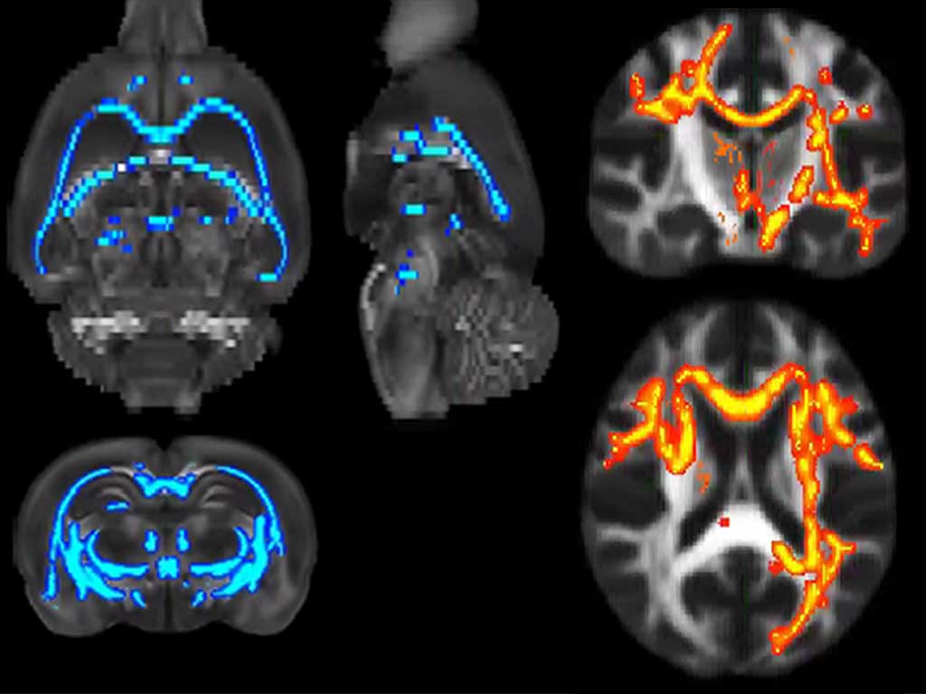
Fractional anisotropy and other DTI measures of white matter properties after long-term alcohol exposure and during early abstinence in both species and clinical and demographic variables and time of abstinence after discharge from hospital in patients.
Results:
The analysis included 91 men with AUD (mean [SD] age, 46.1 [9.6] years) and 27 male rats in the AUD groups and 36 male controls (mean [SD] age, 41.7 [9.3] years) and 9 male control rats. Comparable DTI alterations were found between alcohol and control groups in both species, with a preferential involvement of the corpus callosum (fractional anisotropy Cohen d=-0.84 [P less .01] corrected in humans and Cohen d=-1.17 [P less .001] corrected in rats) and the fornix/fimbria (fractional anisotropy Cohen d=-0.92 [P less .001] corrected in humans and d=-1.24 [P less .001] corrected in rats). Changes in DTI were associated with preadmission consumption patterns in patients and progress in humans and rats during 6 weeks of abstinence. Mathematical modeling shows this process to be compatible with a sustained demyelination and/or a glial reaction.
Conclusions and Relevance:
Using a translational DTI approach, comparable white matter alterations were found in patients with AUD and rats with long-term alcohol consumption. In humans and rats, a progression of DTI alterations into early abstinence (2-6 weeks) suggests an underlying process that evolves soon after cessation of alcohol use.