Español
A translational MRI approach to validate acute axonal damage detection as an early event in multiple sclerosis.
A study from the Institute for Neurosciences detects early axonal damage non-invasively in patients with multiple sclerosis using MRI.
-The results of this work could provide very useful early biomarkers to develop new therapies against multiple sclerosis.
-Axons are the extensions of nerve cells that transmit electrical signals between neurons, allowing them to communicate properly.
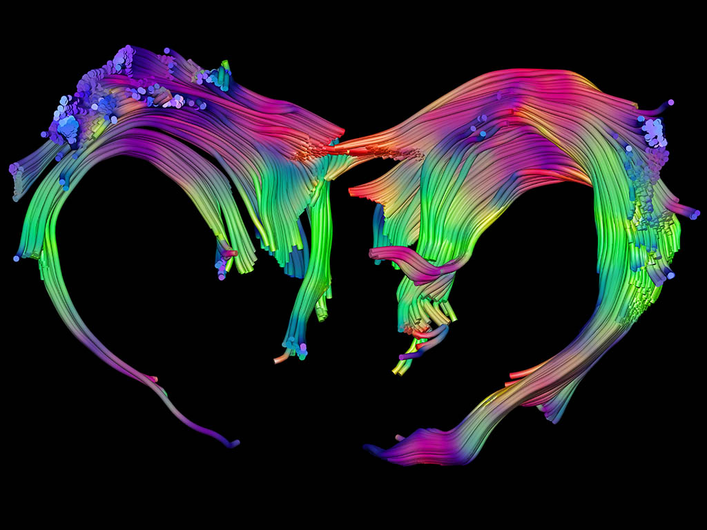
(Photo: Aroa S. Maroto, Silvia De Santis, Antonio Cerdán Cerdá y Jose Antonio Gómez-Sánchez. IN-CSIC-UMH)
Link to Youtube video in the side icon.
To determine whether the increase in axon size can be an early biomarker of multiple sclerosis, the researcher Silvia De Santis, who leads the Translational Imaging Biomarkers laboratory at the Institute for Neurosciences (IN), a joint center of the Spanish National Research Council (CSIC) and the Miguel Hernández University (UMH) of Elche, has directed a joint study with the Athinoula A. Martinos Center for Biomedical Imaging of the Massachusetts General Hospital (Boston, USA) in which they have managed to detect axonal damage in patients with multiple sclerosis in a non-invasive way. To carry out this work, published in the journal eLife, the researchers used the water diffusion-weighted magnetic resonance imaging (MRI) technique.
Millions of people around the world suffer from multiple sclerosis, a chronic, autoimmune disease that affects the central nervous system and whose symptoms range from mobility problems to cognitive difficulties. Although multiple sclerosis primarily affects myelin focally, a crucial, but often least understood, aspect of this disease is diffuse axonal damage, which may be associated with permanent disability. Axons are the extensions of nerve cells that transmit electrical signals between neurons, allowing them to communicate properly. This aspect is essential because when communication between neurons fails, the nervous system cannot develop its functions normally.
Previous research in animal models and post-mortem human tissues suggested that increased axon size could be an indicator of axonal damage, but researchers have had to deal with a great technical challenge: measuring axons in vivo in humans is not possible with traditional imaging techniques. “If we imagine axons as small cables, we must take into account that the diameter of these cables is approximately one micron, hence the complexity of the challenge,” explains Silvia De Santis.
Therefore, researchers have developed an experimental framework to test the ability of water diffusion-weighted magnetic resonance imaging (MRI) to detect the increase in axonal size that is associated with degeneration. This technique has the unique ability to image brain microstructure in vivo non-invasively and with high resolution, by capturing the random movement of water molecules in the brain across different cells and structures.
The researchers confirmed that indeed MRI has the ability to detect changes in the size of axons associated with the acute phase of axonal damage. The first step to verify this was the design of an animal model: “We performed a surgery in which we injected a neurotoxin into a specific area of the rat’s brain and generated an increase in the size of the axons in a controlled manner”, explains researcher Antonio Cerdán Cerdá, first author of the article. The increase in size in the axons was measured by MRI and histological techniques, such as immunohistochemistry or electron microscopy, were used to validate these results.
Finally, to measure axonal size in patients diagnosed with multiple sclerosis, a collaboration was carried out with researchers Caterina Mainero and Nicola Toschi from the Athinoula A. Martinos Center for Biomedical Imaging at Massachusetts General Hospital. In this leading neuroimaging center, affiliated with Harvard Medical School and the Massachusetts Institute of Technology (MIT), researchers were able to use the MRI Siemens Connectom magnet, one of the most powerful and advanced that currently exists worldwide, which allowed them to achieve the necessary sensitivity to carry out the investigation.
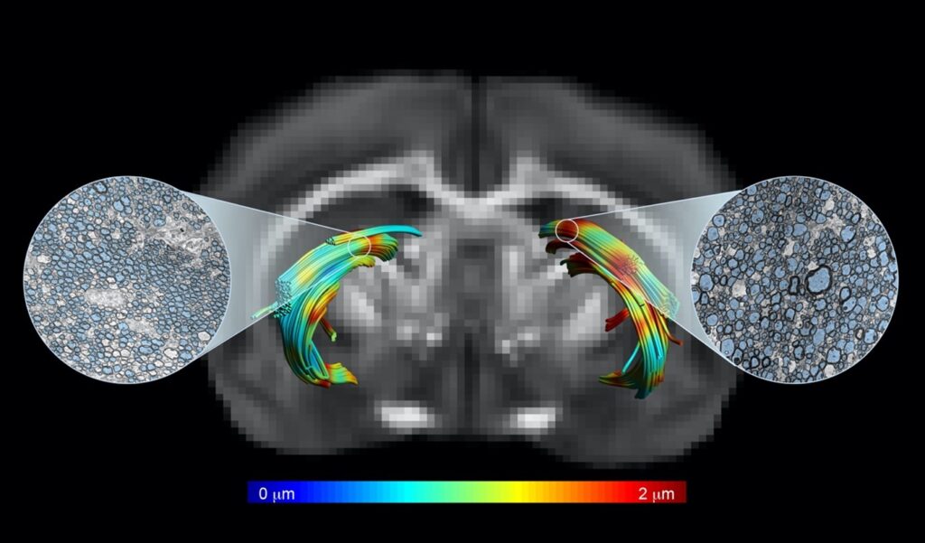 Magnetic resonance image in which two tracts of cerebral white matter have been reconstructed, one with axonal damage (right) and the other healthy (left). Tracts are colored according to axonal diameter measured non-invasively with MRI. Axonal size has been verified through electron microscopy techniques (in blue). IN-CSIC-UMH.
In this procedure, the experts found that there was diffuse axonal damage in most of the white matter of the brain, which is mainly composed of axons and myelin. “The most notable result we found was that the increase in axon size was directly related to early stages of multiple sclerosis disease, confirming our hypothesis that this increase in size may be an early biomarker of the disease,” Cerdán highlights.
Scientific consensus
Until now, some studies argued that water diffusion-weighted magnetic resonance imaging could be capable of measuring axon size non-invasively. However, the lack of validation of this approach generated some controversy in this field of knowledge because some experts considered that this technique did not achieve the necessary sensitivity to measure such a small detail in tissue samples.
Thanks to this study, the researchers have managed to create an experimental framework to test the ability and have reconciled the differences found in previous studies. “Through electronic microscopy techniques, which we have carried out in collaboration with the researcher Jose Antonio Gómez-Sánchez, who directs the Electron Microscopy Platform of the Alicante Health and Biomedical Research Institute (ISABIAL), we have been able to detect that the bias in axonal size that had occurred in previous works is due, at least in part, to the sample preparation process. Therefore, we have confirmed our hypothesis that the axonal size measurements provided by MRI techniques are reliable”, clarifies the researcher.
Impact on patients with multiple sclerosis
The results of this study open the way to the search for new treatments focused on reversing axonal damage. “There are works in animal models of multiple sclerosis that suggest that early axonal damage could be reversible, which reinforces the importance of this finding and raises future avenues for research”, says the researcher, adding that this approach “not only seeks to improve the diagnosis, but to provide a novel tool that will allow in the future to develop new treatments to address the disease from its roots and improve the quality of life of those who suffer from it”.
The laboratory directed by De Santis at the IN-CSIC-UMH focuses on the development, optimization, and application of non-invasive and innovative resonance imaging tools, with a translational approach that is relevant both in basic research and in the clinical setting. Her research pays special attention to healthy aging and pursues the goal of identifying early biomarkers that may precede not only multiple sclerosis but also other neurodegenerative disorders such as Alzheimer’s disease.
This work has been supported by funding from the Spanish Research Agency (AEI) – Ministry of Science and Innovation, the Severo Ochoa R&D Centers of Excellence Program, the Generalitat Valenciana, through the SEJI, CIDEGENT, and ACIF scholarships, the Miguel Servet scholarships program of the Spanish Health Institute Carlos III, and the National Institutes of Health (NIH) of the Department of Health and Human Services of the United States
Source: Institute for Neurosciences CSIC-UMH (in.comunicacion@umh.es)