Multiple parallel cell lineages in the developing mammalian cerebral cortex.
Researchers at the Institute for Neurosciences have discovered the involvement of parallel lineages of stem cells in the generation of neurons.
• This finding represents a significant step towards a greater knowledge of the process of generating neurons, known as neurogenesis.
• Researchers propose that the existence of parallel lineages influences the folding of the cerebral cortex.
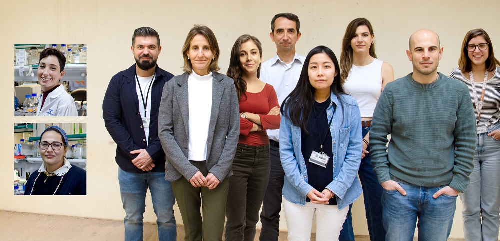
(Photo: IN-CSIC-UMH researchers Virginia Fernández, Salma Amin, Juan Antonio Moreno Bravo, Cristina García Frigola, Lucía del Valle Antón, Víctor Borrell, Yuki Nomura, Ana Prieto Colomina, Adrián Cárdenas and Raquel Murcia Ramón)
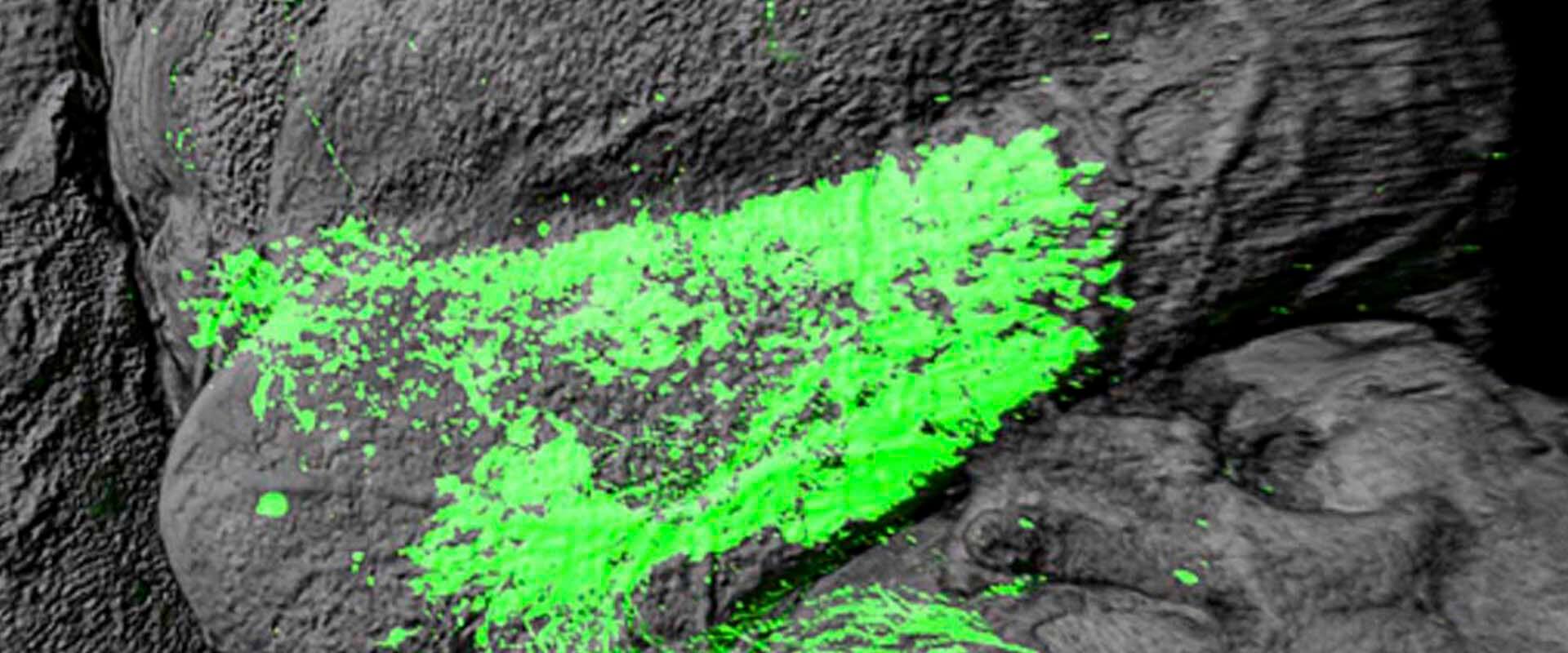
Link to: scFerret developing cerebral cortex
The development of the cerebral cortex largely depends on the stem cells responsible for generating neurons, known as Radial Glial Cells. Until now, it was considered that these stem cells generated neurons following a simple process, that is, a single cell lineage. However, a study led by the Neurogenesis and cortical expansion laboratory, headed by researcher Víctor Borrell at the Institute for Neurosciences (IN), a joint center of the Spanish National Research Council (CSIC) and the Miguel Hernández University (UMH) of Elche, has discovered not only that there are many more types of Radial Glial Cells than previously thought, but also that there are at least three different processes of neurogenesis that occur in parallel in the same brain areas and at the same moment of development.
The results of this work, published in the journal Science Advances, reveal the complexity of neurogenesis through the involvement of parallel lineages. “We have discovered that there are several alternative routes to generate neurons and that all the routes work at the same time, although we have also seen that the final result is always a neuron with similar characteristics and functions at that stage of development”, explains Borrell.
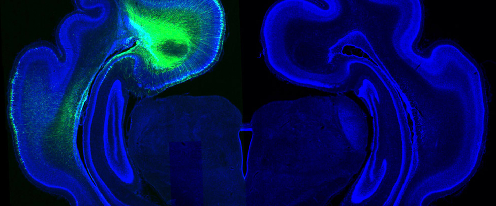
Furthermore, researchers find evidence that the existence of parallel lineages is related to the folding of the cerebral cortex. “A fundamental aspect in this sense is that the ‘routes’ to form neurons work at the same time and in the same place, but not in the same quantity throughout the cortex, being different between gyrus and sulcus”, says the article’s first author, Lucía del Valle Antón. To understand this link, researchers have studied the formation of neurons in regions that will undoubtedly give rise to a gyrus and a sulcus in the ferret brain, while, by using public databases, they have also been able to analyse it in human and mouse brains.
During the development of the study, in which the researcher Juan Antonio Moreno Bravo, who directs the Development, Wiring, and Function of Cerebellar Circuits laboratory, also participated, the experts observed that, although the three lineages are functioning in both gyrus and sulcus zones, different processes predominate depending on the location. “At first, the cortex is smooth, but there is an area that will grow a lot, and as it grows, it will end up forming a gyrus. Meanwhile, next to it, other areas will grow less and will remain sunken, forming a sulcus”, says Borrell, who explains that “The first difference between a gyrus and a sulcus is how much it grows, and this is related to how many neurons will be born in that place. For example, in the sulcus, what we find is that of these three ‘routes’, the one that generates fewer neurons predominates, while in the gyrus, the opposite will happen”.
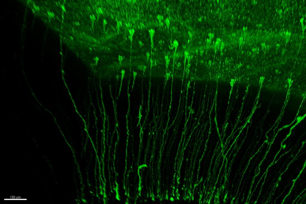
Understanding the existence of these new types of stem cells, which possess a high capacity for division, along with the various mechanisms for generating neurons in parallel, enables us to comprehend the processes that lead to the enlargement of the human cerebral cortex compared to other species.
This research has allowed scientists to explore, with unprecedented detail, the genes expressed by neurons in both the gyrus and the sulcus. Víctor Borrell explains: “We aimed to observe which of all the cells we investigated express genes known to be mutated in human malformations. We verified that not all these cells express the genes responsible for these brain malformations. We observed that they are mainly expressed by the newborn neurons, rather than the progenitors”.
Along these lines, the researcher highlights that, despite having the same functions at a global level, the neurons that are born in the gyrus express genes that are essential for the human cortex to have gyri. This indicates that, when patients have malformations because their brain lacks gyri, the defects occur specifically in the neurons of the gyrus and not in those of the sulcus.
International collaboration
In this study, which involved collaboration with researchers from the ISF Stem Cell Research Institute (Helmholtz Zentrum) and the Max Planck Institute for Biological Intelligence, both located in Munich (Germany), the researchers based their results on the sequencing of individual cells at the transcriptomics level, a technique that enables us to identify all the genes that are expressed in each of the cells.
Scientists analysed thousands of cells using informatics tools to determine the genetic trajectory of these cells and their respective lineages. Upon investigating and validating the lineage data across the three species, they observed that in the human brain, these three parallel lineages also occur, similar to what is observed in ferrets. However, in the case of mice, analyses conducted have observed only a single predominant route in the creation of neurons. Future research will be necessary to determine whether mice lost these lineages due to evolution or if, on the contrary, these ‘routes’ are still present but in such small proportions that they are undetectable with current tools.
This work has been made possible thanks to funding from the Spanish State Research Agency – Ministry of Science, Innovation, and Universities through the Generación de Conocimiento, FPI, and Juan de la Cierva programs, the Severo Ochoa program for centers of excellence, and the Tatiana Pérez de Guzmán el Bueno Foundation.
Source: Institute for Neurosciences CSIC-UMH (in.comunicacion@umh.es)

 Español