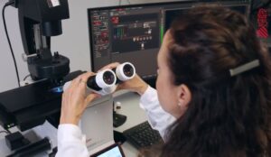
Eye protection device against infections intended to be placed in the eyepieces of a microscopy equipment [U202032487]
Inventors: Víctor Rodríguez, Joana Expósito and Verona Villar
Type: Utility model
Date: 2020
Technology

Microscopy systems are generally shared use, mainly due to their high cost and the large space they require. The shared use of microscopy equipment can cause the transmission of infections through the ocular conjunctiva from one person to another. In order to avoid the transmission of diseases through this route, many research centers have implemented different prevention measures, among which are the continuous disinfection of the eyepieces with ethanol, the mandatory use of safety glasses or the coating of the eyepieces. eyeglasses with film prior to each use. However, these measures have not turned out to be the definitive solution to the problem, since all of them have some drawback. On the one hand, the repeated use of ethanol as a disinfectant causes the deterioration of the oculars, on the other hand, the use of glasses and/or film to cover the eyes and/or oculars greatly reduces the quality of the visualization of the samples. . Currently, a completely effective solution to this problem is not known in the state of the art, so, in view of this situation, research technicians from the Institute of Neurosciences CSIC-UMH have developed an eye protection device against infections to be placed in the eyepieces of microscopy equipment. This new device for individual use allows personal protection in the use of any microscopy system in an efficient, simple and economical way.
Benefits
- Reduction of the risk of contagion of infections through the ocular conjunctiva caused by the shared use of microscopy equipment.
- Device easy to use, store and transport.
- Comfortable use and quality vision.
- Cost savings due to the simplicity of the device, economic and large-scale manufacturing.
- Device for individual use with a long useful life, admits cleaning and disinfection, as well as the replacement of the transparent sheet in case it is damaged by use.
- Scalable and adaptable to the size of the eyepieces of different equipment.
- Avoid the generation of large amounts of plastic.
- Prevents wear of the eyepieces by the action of disinfection products.
State of development
There are 300 units (150 pairs) of the device prototype manufactured using 3D printing technology. They are currently being used satisfactorily by all users of microscopy equipment at the Institute of Neurosciences, in microscopes and magnifying glasses from different commercial houses: Leica, Zeiss, Olympus and Nikon. The device has been protected by means of a utility model submitted to the Spanish Patent and Trademark Office. Currently, a mold for its manufacture has not been produced, however, 3D designs are available for its realization. Likewise, its manufacture through molds is recommended, in order to lower costs and reduce manufacturing time.
Collaboration
The IN seeks a collaboration that leads to the commercial exploitation of the invention presented. The best scenario for the institution would be to reach an agreement to transfer the use of the technology by sale or license (exclusive or non-exclusive) of the technology presented. However, the form, terms and conditions of collaboration can be openly discussed if the technology presented is of interest.
pdf document
Surface antenna on sample positioning device [P202130726]
Inventors: Luis Tuset and Víctor Rodríguez
Type: Patent
Date: 2021
Technology
Nuclear magnetic resonance (NMR) is an imaging technique widely used in current biological research that combines the application of a strong magnetic field and an array of coils and antennas to detect a radiofrequency signal from the sample. There are three types of antennas to use depending on the area of interest to study, such as volume, internal or surface. Surface antennas are widely used as they provide a high signal-to-noise ratio; however, there are some drawbacks to keep in mind about them. The existing positioning techniques that are currently used are insufficient and imprecise, they consist of the use of adhesive tape to fix the antenna to the sample. Also, the antenna must be connected to a preamplifier via a cable that can be bent depending on the position of the antenna, creating more problems in image acquisition. Researchers from the Institute of Neurosciences of the Miguel Hernández University have developed a solution that consists of a positioning device that offers a precise technique for mechanical placement of the antenna. The device is based on the positioning of the antenna by means of a rotating arm connected to a chassis, which allows obtaining better quality results in preclinical studies of small animals.
Benefits
- The device design achieves a more precise, flexible, and replicable array of the antenna, allowing closer positioning to the area of interest.
- With the mechanical arm in charge of placement, involuntary displacements cannot occur, which avoids having to repeat the test after a first attempt.
- The design of the device allows the combination of brain studies with stimulation techniques. In addition, surgical procedures can be performed at the time of image acquisition.
- The device prevents the bending of the cable which eliminates the associated interference.
State of development
The benefits have been extensively tested in combination optogenetic/fMRI experiments in awake rats and calibration experiments in MRI phantoms. A Spanish patent application has been filed with the Patent and Trademark Office (OEPM) in July 2021.
Collaboration
The IN seeks a collaboration that leads to a commercial exploitation of the present invention. The ideal scenario for the institution would be to reach an agreement to transfer the technology, use by sale or license (exclusive or non-exclusive). However, the form, terms, and conditions of the collaboration can be openly discussed if the presented technology is of interest.
Pdf DocumentFlyGear: Automatic and accurate monitoring of insects [PCT/ES2020/070670]
Inventors: María Domínguez
Type: Patent
Date: 2020

The use of Drosophila melanogaster in research is widespread in sectors such as pharmacology and biotechnology, and its use as a biofactory is gaining interest, being the main model organism for high-throughput studies of drugs in cancer, longevity, and neurodegeneration with resolution of the disease. population and individual behavior, in which development time is a key factor for predicting toxicity and disease. The Institute of Neurosciences (CSIC and UMH) has developed a robot and software that allows counting and measuring, in an automated, precise and reliable way, the transition time between vital phases of the fruit fly (Drosophila melanogaster) and other insects. The precise measurement of the time of transition to adulthood is essential in numerous investigations on cancer and longevity, detection of therapeutically useful compounds, and scientific studies in developmental biology on the impact of hormones or environmental factors. Due to its robust and compact design, the device can be housed in most incubators and used with other sensors for temperature, humidity, etc. allowing absolute precision and performance.
Benefits
- It incorporates a light source (visible and infrared LEDs) to obtain high-quality images, and for studies of the circadian cycle.
- Allows you to configure the image-taking time and lighting conditions to your liking, with easy-to-use software.
- Data collection includes: time, number of individuals, size, life stage, etc. It allows the storage and processing of data in the cloud.
- Portable system and compatible with most fly containers. It can be adapted to other species of flies and insects.
- The device has been designed to meet the need to scale this task to the industrial level for the biotech and pharmaceutical industries.
- Artificial intelligence system for automatic recognition of all fly development milestones.
State of development
The PCT has now entered the national phases in the United States and Europe.
Collaboration
The IN is looking for pharmaceutical companies and/or laboratory equipment manufacturers interested in the patent license for the industrialization and commercialization of the device.
PDF document

 Español