Español
Thalamic Activity Regulates Interneuron Density in the Developing Visual Thalamus.
Study reveals that the thalamus actively contributes to the development of visual circuits
• A study by the Institute for Neurosciences CSIC-UMH reveals that thalamic activity regulates the integration of interneurons in a key region of the visual pathway.
• The results, obtained in mouse models, show that altering this activity also affects the organization of these cells in the visual cortex.
(Photo: From left to right: IN CSIC-UMH researchers Pablo Castellano-Ruiz, Daniel Torres-Romero, Emily Wilson, Francisco José Martini, and Guillermina López-Bendito)
A team led by researcher Guillermina López-Bendito at the Institute for Neurosciences (IN), a joint center of the Spanish National Research Council (CSIC) and Miguel Hernández University of Elche (UMH), has shown that the thalamus’s own activity plays a fundamental role in the organization of visual circuits during development. Published in The Journal of Neuroscience, the study shows in mouse models that altering this activity changes the number and distribution of interneurons — neurons that help regulate the activity of neural circuits — in the visual thalamus, and also affects the organization of these cells in the visual cortex.
The thalamus plays a central role in communication between the sensory organs and the cerebral cortex. In vision, it receives information from the retina and transmits it to the visual cortex, but the results of this study show that during development it also plays a role in organizing the circuits that will process this information.
“These results show that the thalamus does not simply act as a relay for visual information, but also plays an active role in the development of neural circuits. Its activity helps regulate how interneurons are incorporated and distributed, a process that is essential for these circuits to develop properly”, explains Guillermina López-Bendito, who leads the Development, Plasticity and Reprogramming of Sensory Circuits laboratory at the IN CSIC-UMH.
The brain begins to organize its circuits before vision begins
The development of the visual system begins long before animals can receive visual information from their surroundings. During the early stages of brain development, neurons generate spontaneous activity that helps organize neural circuits. In mice, many of these processes take place before the eyes open.
To study the role of this activity, the team used different mouse models in which they could alter neuronal activity at different points along the visual pathway and at different stages of development. In one of the models, the researchers were able to modify thalamic activity without eliminating the connections between the retina and the thalamus.
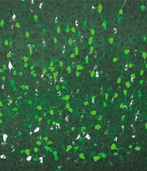
Interneurons in the visual cortex, one of the cell populations whose organization is influenced by thalamic activity. Author: Irene Huerga-Gómez.
“The important thing is that we have been able to separate the contribution of the retina from the thalamus’s own activity. By specifically altering thalamic activity, we observed changes in the density and proportion of interneurons in the visual thalamus”, says Pablo Castellano-Ruiz, co-first author of the study together with researchers Irene Huerga-Gómez, Daniel Torres-Romero and Emily Wilson.
A necessary ‘brake’ for neural circuits
The study focuses on inhibitory interneurons, a group of cells whose function is to regulate the activity of other neurons. “In simple terms, these cells act as a brake that prevents neural circuit activity from becoming uncontrolled and help maintain the conditions required for the proper functioning of neural networks”, explains López-Bendito.
The researchers analyzed the number, density, and distribution of these interneurons in the dorsal lateral geniculate nucleus, a region of the thalamus that is part of the visual pathway. The results showed that intrinsic thalamic activity is a key factor in regulating the density of these cells: when this activity is altered during development, their proportion in this region increases. This effect is not explained by increased interneuron production or survival, but rather by changes in their integration and distribution within the circuit.
The experiments also made it possible to distinguish this mechanism from the role of the retina. While the absence of retinal connections mainly alters the positioning of interneurons in the thalamus, changes in intrinsic thalamic activity affect their proportion. Thus, both systems contribute to the organization of the visual circuit during development through different yet complementary mechanisms.
In addition, the researchers found that altering thalamic activity during prenatal development affects the organization of interneurons in the visual cortex. Changes were observed across different populations of these cells and, in some cases, persisted into later stages of development, indicating that thalamic activity can influence the organization of circuits beyond this structure.
This work was made possible thanks to funding from the European Research Council under the European Union’s Horizon 2020 Research and Innovation Programme (ERC Advanced Grant SPONTSENSE and ERC Consolidator Grant SENSORTHALAMUS); the Spanish Ministry of Science, Innovation and Universities; the “Severo Ochoa” Programme for Centers of Excellence of the Spanish State Research Agency; and the PROMETEO programme of the Generalitat Valenciana.
Source: Institute for Neurosciences CSIC-UMH (in.comunicacion@umh.es)