Español
GluN3A Is Required for Coordinated Postnatal Development of Axonal and Dendritic Branching Patterns in Mouse L2/3 Callosal Projection Neurons
Study reveals that a protein coordinates when and where connections between the two brain hemispheres are formed
• The study, led by the Institute for Neurosciences, reveals that GluN3A synchronises neuronal maturation with the pattern of connections they receive during the early stages of life.
• In the absence of GluN3A, dendritic trees branch prematurely, while axons from the opposite hemisphere are distributed across different regions of the cerebral cortex.
(Photo: Researchers Bárbara Corral Sánchez, Isabel Pérez Otaño and Ana Isabel Navarro at the Institute for Neurosciences CSIC-UMH. Source: IN CSIC-UMH.)
During brain development, neurons form an enormous number of connections. But building a functional circuit requires more than one neuron simply making contact with another: connections must form at the right time and occupy specific positions within the cells that receive them. In this context, a study led by researcher Isabel Pérez Otaño at the Institute for Neurosciences (IN), a joint centre of the Spanish National Research Council (CSIC) and Miguel Hernández University of Elche (UMH), has identified a mechanism that helps coordinate when and where certain connections between neurons are established during brain development.
The results, published in The Journal of Neuroscience, show that the protein GluN3A acts as a regulator of neuronal connection maturation, preventing some of their components from developing too early. Neurons communicate with one another through specialised extensions: axons transmit signals to other cells, while dendrites branch out to receive them, forming extensive ‘dendritic trees’. When this protein is absent, the dendrites of certain cortical neurons branch prematurely, while axons arriving from the opposite hemisphere alter their distribution, disrupting the precise organisation of the circuit.
To study this process, the team used mouse models that allow them to visualise the development of neurons and their connections during the early stages of life. “GluN3A acts as a brake during a specific stage of development, preventing certain synaptic connections from maturing too early or excessively”, explains Pérez Otaño, who leads the Plasticity and Remodeling of Neural Circuits laboratory at the IN CSIC-UMH.
To understand where GluN3A acts, the team analysed separately the neurons that send the connections and those that receive them. Using three-dimensional reconstructions of individual neurons, they were able to study the shape and distribution of their dendrites in detail. The experiments showed that the effect occurs mainly in the neurons that receive the connections, rather than in those that send them.
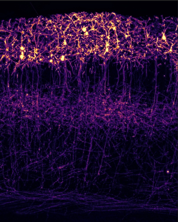
Image of the somatosensory cortex of a mouse, showing neurons in the superficial layers, including their cell bodies, dendrites, and axonal projections. Selected as the cover image for the issue of The Journal of Neuroscience, the image illustrates the role of GluN3A in organising these connections during brain development. Credit: Bárbara Corral Sánchez.
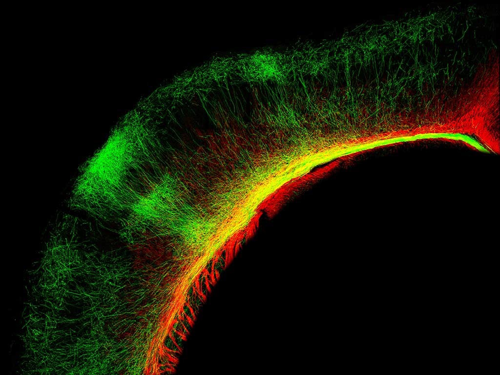
The researchers also observed that, in the absence of GluN3A, the axons connecting the two brain hemispheres are not distributed in the same way as in control animals. Although these axons are able to reach the opposite hemisphere and cross the corpus callosum, they subsequently reach different areas within the cortex. This indicates that the alteration is not due to a problem in reaching the opposite hemisphere, but rather to a subsequent change in the environment they encounter.
“When this key protein is absent, dendrites become more complex too early, but this does not necessarily mean that the connections are formed more effectively,” explains Bárbara Corral Sánchez, co-first author of the study together with Ana Isabel Navarro and Oliver Crawley. “The development of a connection depends not only on neurons growing, but also on their different components developing in a coordinated way”, adds the researcher.
Molecular mechanisms
To investigate further the mechanisms that could explain these changes, Pérez Otaño’s team analysed gene activity in neurons from mice with and without GluN3A. The analysis identified differences in the expression of genes involved in dendritic development and axonal growth, including CRMP4 and KCNA1.
The results suggest that the absence of GluN3A alters cellular programmes involved in the maturation of different components of the neuronal circuit, and that this process is necessary for connections to be established with precision. This new information could help explain why genetic mutations in GluN3A are associated with neuropsychiatric disorders in which neuronal connectivity is affected.
The study was conducted in mouse models and during early developmental stages, so its results provide insight into basic mechanisms involved in the organisation of neuronal circuits. “It is essential to understand how neuronal circuits are built from the beginning, because understanding how they are organised and mature allows us to better understand how the brain works and what can happen when these processes are disrupted”, says Pérez Otaño.
This work was possible thanks to the funding from the PROMETEO Programme of the Generalitat Valenciana; the Spanish State Research Agency – Ministry of Science, Innovation and Universities; and the Severo Ochoa Programme for Centres of Excellence. The study also received support from predoctoral fellowships from the Generalitat Valenciana (CIACIF/2021/453), Marie Curie (H2020-MSCA-IF-2019) and CSIC (IFERC20007).
Source: Institute for Neurosciences CSIC-UMH (in.comunicacion@umh.es)