Español
Callosal inputs generate side-invariant receptive fields in the barrel cortex.
A study from the Institute for Neuroscience CSIC-UMH describes how sensory information is transmitted between brain hemispheres
• One more step to understand how the brain processes sensory information.
• Researchers have described for the first time how the mirror effect is produced giving continuity to the receptive field of both hemispheres and making them act as one.
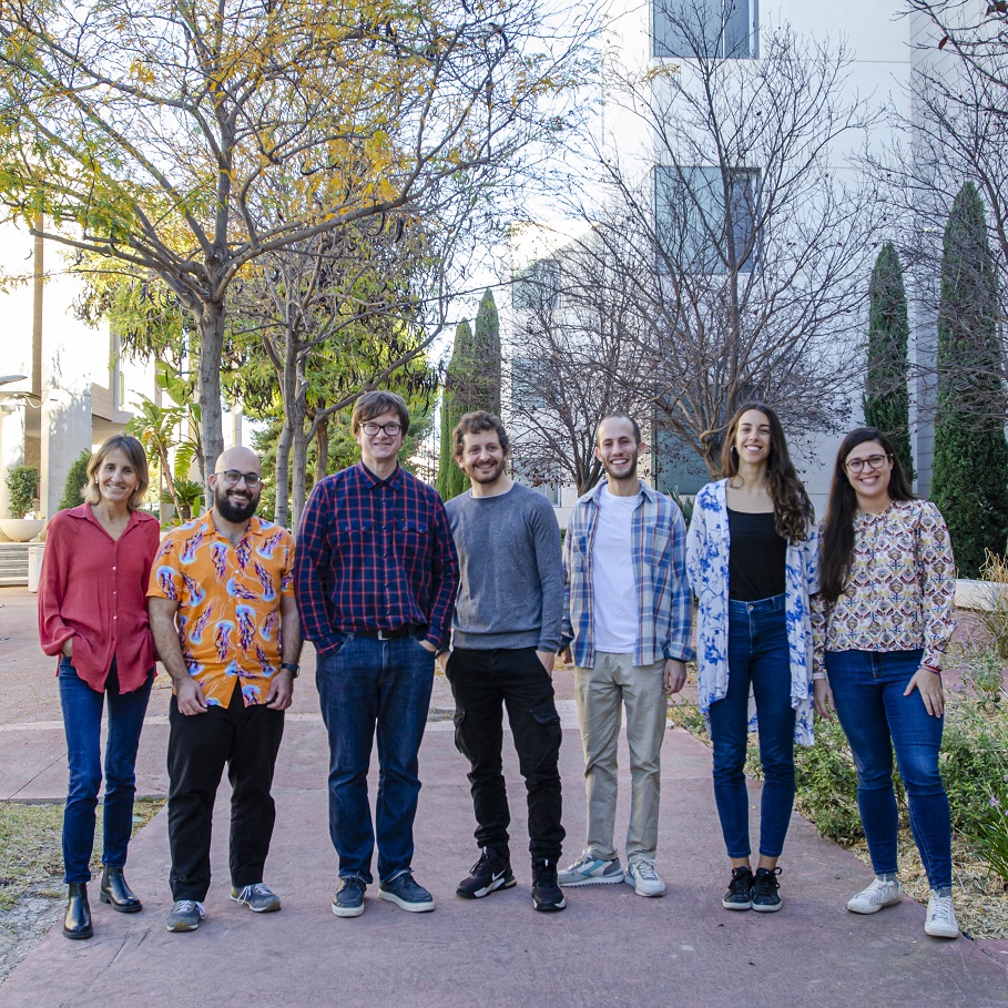
(Photo: Cristina García, Javier Alegre, Ramón Reig, Roberto Montanari, Ismael Navarro, Alicia Alonso & María Sáez, IN-CSIC-UMH researchers)
The brain has a very specific way of processing information from the sense of touch. Stimuli felt on the left side of the body are processed in the right brain hemisphere and vice versa. This is a widely known issue, but until now it has not been described in detail how the two hemispheres share this information. Researchers from the Sensory-motor processing by subcortical areas laboratory, led by Ramón Reig at the Institute for Neurosciences (IN), a joint center of the Spanish National Research Council (CSIC) and the Miguel Hernández University (UMH) of Elche, have carried out a study that confirms, in a pioneering way, through physiological studies, that a double representation occurs between hemispheres, which allows the perception of continuity, without interruptions between both sides of the body.
This work, published in the journal Science Advances, addresses a hypothesis known as Midline Fusion Theory. This theory was postulated in 1989 and, based on anatomical observations, it established that the areas of the brain that encode sensory information close to the midline of the body sent a large number of connections that crossed to the other hemisphere. Until now, the presence of these axons has been observed, but IN researchers have demonstrated in mice the functional properties of those axons that cross and synapse with the other hemisphere. They especially connect tactile information from the midline parts and generate an identical representation or activation of the information on both sides, allowing sensory information to be processed continuously.
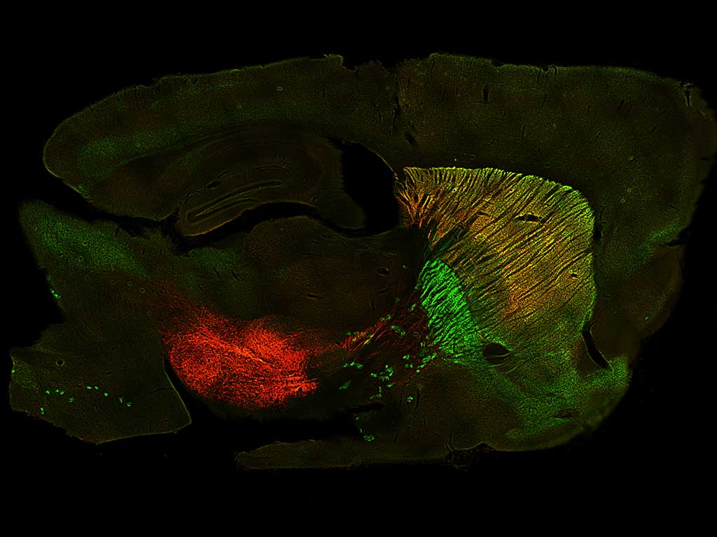
To carry out this study, the researchers have used the mouse whisker system, since these rigid hairs are excellent tactile receptors that allow both the lateral areas and the midline areas to be stimulated with great precision. In addition, it is possible to observe in detail which part of the hemisphere responds to the stimulus. The experts were able to verify that when a certain whisker on the mouse’s snout close to the midline is stimulated, not only is a response produced in a specific contralateral location, but a third of the neurons in the homologous region of the opposite hemisphere respond exactly the same. This explains how the brain is able to generate that tactile spatial continuity between both sides of the body.
Furthermore, thanks to the neuron recording technique used (in vivo patch-clamp), the researchers also verified that, when the tactile response occurs, in the opposite hemisphere (ipsilateral to the tactile stimulation) through the neurons that cross the corpus callosum, it receives not only the response that causes the activation of the neurons but, a few milliseconds later, it also receives the inhibition necessary to control the response. The correct balance of excitation-inhibition of neurons is essential for the brain to develop its activity normally since a continuous state of excitation would trigger an epileptic brain.
In this work, whose first author is Roberto Montanari, they have managed to precisely describe the complete circuit that connects both cerebral hemispheres: the information perceived in response to a sensory stimulus travel through the corpus callosum and is specifically processed in a very specific region of the primary somatosensory cortex, in the mouse the barrels of row A. Therefore, these represent a sensory center for interhemispheric communication.
Furthermore, they have verified that information travels through a specific ‘lane’. The cerebral cortex that encodes the mouse’s tactile information is divided into rows and columns, each containing groups of neurons called barrels. Researchers have proven that communication between hemispheres occurs in row A: “This is what is called heterotopic projection. For example, the barrels in row E barely project to row E of the other hemisphere, but rather do so mostly through row A”, explains Ramón Reig and adds that this is very interesting because it is precisely in row A where the midline whisker receptors are located.
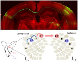
Above: Neurons from one hemisphere of the primary somatosensory cortex project to the other hemisphere. Below: Scheme showing how information travels between hemispheres once stimulation occurs. Neurons in row A barrels contact adjacent barrels and row A in the contralateral hemisphere. IN-CSIC-UMH
The experts used a common anesthetic (lidocaine) to block all information coming from one side of the mouse’s snout in order to see what happened when the animal could only process information from one side. Once again, they verified that the information travels through row A connecting both hemispheres.
To validate these results, the researchers imitated what they had carried out at the sensory level in the mouse whiskers with optogenetic techniques. The experiment consisted of directly stimulating the cerebral cortex with light to observe the response of the neurons in rows A and, indeed, they observed that the response coincided and gave rise to the same phenomenon.
One step further: the striatum
Researchers have discovered that the dorsolateral striatum not only receives tactile information but also processes tactile information bilaterally, from both hemispheres. The laboratory directed by Reig at the IN studies the striatum to understand how the neurons in this brain region integrate sensory and motor information to produce a coordinated and precise response. Problems in the function of this nucleus are related to motor disorders such as Parkinson’s disease. This new research also precisely describes the route that bilateral tactile information follows before reaching the striatum.
This work has been possible thanks to funding from the Ministry of Innovation, Science and Universities, the CSIC Severo Ochoa Excellence Program of the Institute for Neurosciences, la Caixa, and the ACIF Program of the Generalitat Valenciana.
Source: Institute for Neurosciences CSIC-UMH (in.comunicacion@umh.es)