Español
Brain dynamics supported by a hierarchy of complex correlation patterns defining a robust functional architecture.
A study co-led by the Institute for Neurosciences reveals the brain’s dynamic architecture using advanced MRI techniques.
– Experts have incorporated communication delays between brain regions, allowing them to discover a robust and dynamic brain architecture.
– This work paves the way for identifying more precise and sensitive brain biomarkers.
(Foto: Santiago Canals, investigador IN, CSIC-UMH)
Researchers from the Institute for Neurosciences (IN), a joint center of the Spanish National Research Council (CSIC) and the Miguel Hernández University (UMH) of Elche, along with a team from the Transylvanian Institute of Neuroscience (Romania), and in collaboration with experts from the Universitat Politècnica de València, have developed an innovative approach to studying brain connections using functional magnetic resonance imaging (fMRI). This work, recently published in the journal Cell Systems, introduces a new way of understanding brain architecture through dynamic functional networks, challenging the traditional static approach.
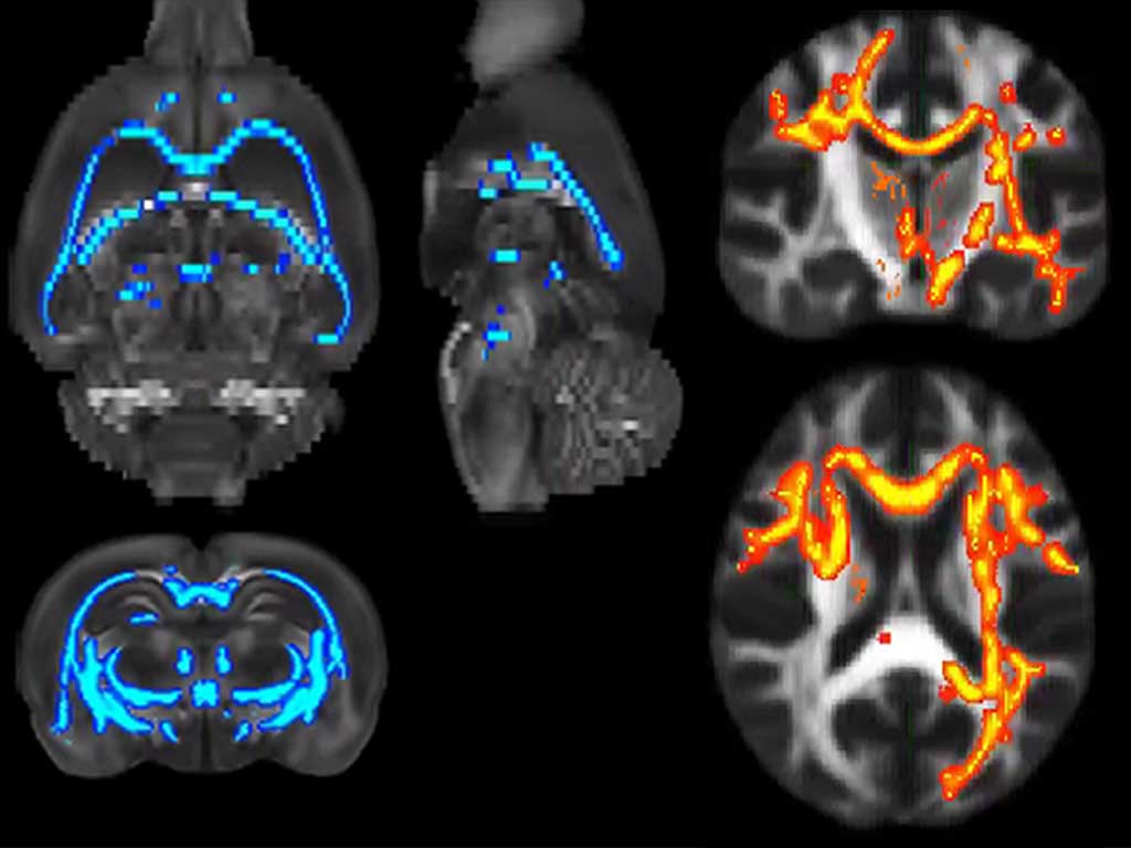
The study has revealed that delays in communication between brain regions are key to understanding the organization of functional networks. Unlike conventional methods that average a single static network, the team used an approach that studies the temporal evolution of connection strength, analyzing their statistical distribution rather than their average activation. This has allowed them to discover a brain architecture that is both robust and dynamic.
“The connection speed between brain regions and integration times are variable, which introduces different communication delays. Our objective has been to incorporate these delays into the analysis of functional connectivity to develop a more precise and sensitive method”, explains researcher Santiago Canals, who leads the Plasticity of Brain Networks laboratory at the IN.
The results of this research reveal the existence of a functional backbone formed by robust, delay-free interactions, complemented by a large number of weaker connections whose strength fluctuates over time, adding flexibility to the brain’s functional architecture. “This dynamic approach better captures the brain’s constantly changing reality. It has enabled us to obtain comparable results in rats, marmosets, and humans, with extraordinary consistency when the same subject is scanned repeatedly over time, which is uncommon in the field of magnetic resonance,” Canals emphasizes.
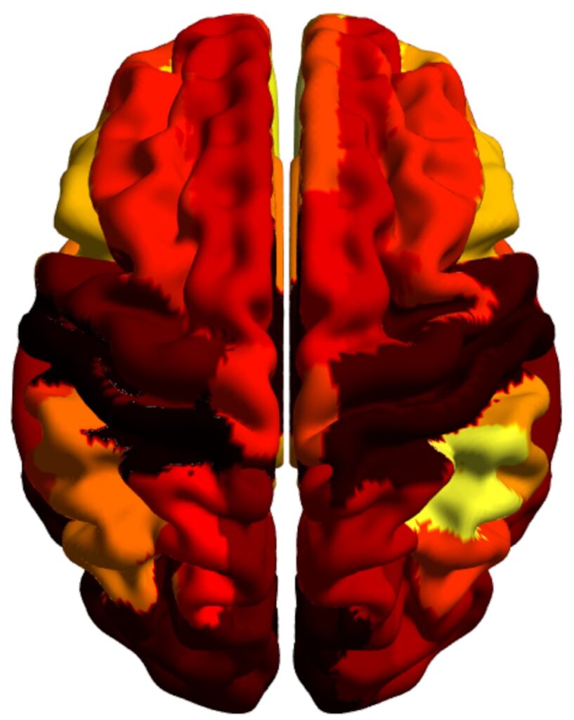
The image shows the hierarchical distribution of connections represented in colors on the surface of a human brain. (Credits: Cell Systems)
One of the most significant discoveries of the study is the identification of the “backbone” — a set of extremely strong and stable functional connections that serve as the core of communication in the brain. Although these connections represent less than 10% of all those studied, they play a crucial role in the overall cohesion of brain networks, maintaining robust connectivity that ensures efficient communication between different regions. “Efficiency in network communication is dramatically reduced when these connections are compromised, underscoring their importance in the brain’s functional structure. Meanwhile, the weaker and more dynamic connections greatly amplify the system’s potential functional states, providing flexibility”, concludes Canals.
To conduct this study, scientists used fMRI data from rats, non-human primates, humans, and patients with alcohol use disorder. These findings open new avenues for identifying more precise and sensitive brain biomarkers capable of detecting subtle alterations in neuronal networks, which could have significant implications for diagnosing neuropsychiatric diseases.
These results were made possible through a joint international collaboration that brought together experts from various fields. Canals emphasizes that the research would not have been possible without a multidisciplinary team. The laboratory led by Maria Ercsey-Ravasz at the Transylvanian Institute of Neurosciences (TINS) has extensive experience in the physics of complex networks, while Raul C. Muresan’s lab at the same institution focuses on developing advanced tools for time series analysis. Additionally, David Moratal’s group from the Center for Biomaterials and Tissue Engineering (CBIT) at the Universitat Politècnica de València also contributed to the study.

(Photo: Mohamed Kotb Selim, Laura Pérez-Cervera and Santiago Canals, IN-CSIC-UMH researchers)
This work has been possible thanks to the funding of the European Union’s Horizon 2020 Research and Innovation Programme, the ERA-Net NEURON Programme, the Spanish Agency of Research (AEI), the European Regional Development Fund (ERDF), the Spanish Ministry of Health, Social Services and Equality, the PROMETEO program of the Generalitat Valenciana, the Severo Ochoa Programme for Centres of Excellence in R&D, the Human Brain Partnering Project ERANET-FLAG-ERA-ModelDXConsciousness and ERANET-NEURON-2-UnscrAMBLY project, the National Authority Romanian Society for Scientific Research and Innovation, and the German Research Society (DFG).
Source: Institute for Neurosciences CSIC-UMH (in.comunicacion@umh.es)