A Proteomic Analysis Reveals the Interaction of GluK1 Ionotropic Kainate Receptor Subunits with Go Proteins
Kainate receptors (KARs) have the structure of ionotropic glutamate receptors, but unlike AMPA and NMDA receptors, KARs can activate metabotropic signalling (non canonical signalling) as well as passing ionic current (canonical). Several metabotropic effects of KARs have been demonstrated, including inhibition of voltage-sensitive calcium channels, inhibition of glutamate and GABA release, and inhibition of the slow afterhyperpolarization current (IAHP). All these effects are blocked by pertussis toxin and involve phospholipase C, suggesting they are mediated by Go proteins, but how KARs activate G-proteins has remained an open question.
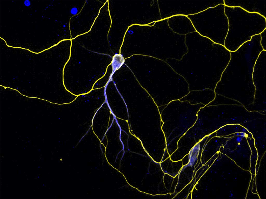
To answer this question, Rutkowska-Wlodarczyk et al. performed a proteomic analysis on mouse brain homogenates and identified proteins that interacted with the intracellular C-terminal portion of GluK1b, a KAR subunit. They found 22 proteins that specifically interacted with GluK1b, including the α subunit of Go. When coexpressed in neuroblastoma cells, GluK1b and Gαo were partially colocalized. Furthermore, fluorescence was reconstituted when cells were transfected with the C- and N-terminal fragments of yellow fluorescent protein fused to Gαo and GluK1b, respectively, indicating that Gαo and GluK1b are closely apposed in vivo. Additional experiments using bioluminescence resonance energy transfer demonstrated that glutamate activated Go in GluK1b-expressing HEK cells. Finally, kainate-induced reduction of IAHP in dissociated mouse dorsal root ganglion (DRG) neurons was abolished by knocking out GluK1, but not by knocking out GluK5, the only other KAR subunit expressed in these cells.
These data strongly suggest that direct interaction between GluK1b and Gαo underlies KAR on canonical signalling further clarifying mechanisms of synaptic transmission and modulation mediated by these elusive glutamate receptors.

 Español
