
 Español
Scientists find a determining mechanism for the synapse formation in the cerebellum
9 de July de 2024
- A study co-led by the Institute of Neurosciences (CSIC-UMH) demonstrates that one of the receptors for the neurotransmitter glutamate is essential to generate synaptic plasticity in the cerebellum.
- Dysfunctions in synapses can lead to brain disorders such as epilepsy, Parkinson's disease, depression, schizophrenia, and autism.
Kainate receptors regulate synaptic integrity and plasticity by forming a complex with synaptic organizers in the cerebellum. Kakegawa W, Paternain AV, Matsuda K, Aller MI, Iida I, Miura E, Kazuya Nozawa K, Yamasaki T, Sakimura K, Yuzaki M and Lerma J. Cell Reports, 2024.
DOI: https://doi.org/10.1016/j.celrep.2024.114427

(Photo: María Isabel Aller, Ana Valero Paternain y Juan Lerma, IN-CSIC-UMH researchers)
The Institute of Neurosciences (IN), a joint center of the Spanish National Research Council (CSIC) and the Miguel Hernández University (UMH) of Elche, leads together with Keio University in Tokyo (Japan) a study that demonstrates the crucial role of one of the receptors for the neurotransmitter glutamate in the functioning of the synapses of the cerebellum. In a work published today in the journal Cell Reports, they describe the molecular mechanism by which kainate receptors not only act as synaptic receptors, but also as 'scaffolds' that support the structure of connections between neurons. These results enable the design of new synaptic connectors using specific combinations of kainate receptor subunits and offer promising avenues for future therapeutic applications.
Synapses are the connection points where neurons establish contact with each other to transmit information through nerve impulses. For this communication to occur, the presynaptic neuron releases a neurotransmitter, which is then received by a postsynaptic neuron. The Synaptic Physiology laboratory directed by CSIC researcher Juan Lerma at the IN has extensively investigated glutamate receptors, which are neurotransmitters involved in various processes of the central nervous system, particularly kainate receptors, one of the three families of glutamate receptors that mediate communication between neurons. "For many years, we have tried to find out what the function of kainate receptors is in synaptic physiology and brain pathologies", says the researcher.
His laboratory has made significant contributions to understand the roles of these proteins in synaptic communication, which, when dysfunctional, lead to multiple neurological and neuropsychiatric disorders. This team of IN researchers had previously detected the role that the GluK4 protein, one of the five subunits that make up kainate receptors, can play when it is overexpressed in pathologies such as autism, depression, and anxiety. They also demonstrated that the GluK1 protein is triplicated in patients of Down Syndrome and that these decompensated levels are responsible for the spatial memory deficits observed in these patients.
Furthermore, the laboratory directed by Michisuke Yuzaki in the Department of Neurophysiology at the Keio University School of Medicine in Tokyo has studied the functioning of synapses in the cerebellum over the years and discovered that an interaction between the C1ql1 and Bai3 proteins occurs in this region to enable the formation of synapses. However, the results of this new study modify this concept by demonstrating that without the interaction of both proteins with kainate receptors, synapses do not form: “By combining our experience and knowledge in this new collaboration, we have been able to completely redefine synapse formation in the cerebellum” highlights Yuzaki.
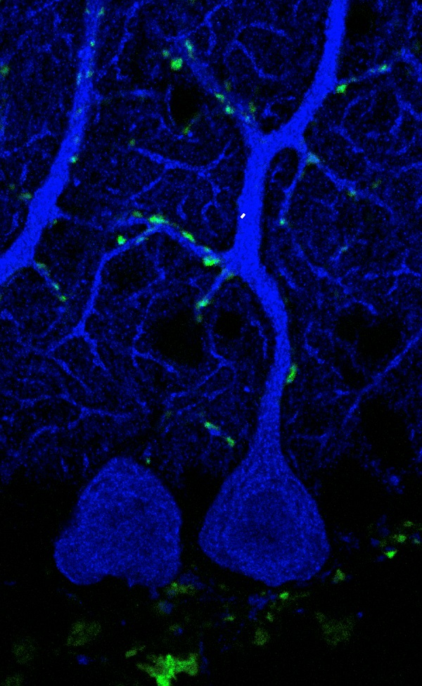
Expansion microscopy image with synaptic boutons (green) of climbing fibers on a Purkinje cell (blue) from the cerebellum of a mouse. Credits: IN (CSIC-UMH)
The experts confirmed that the presence of GluK4, which is expressed by cerebellar Purkinje neurons, is essential for the interaction that supports synaptic transmission between climbing fibers and these neurons to occur. To confirm this, the researchers used mouse models in which they genetically manipulated the expression of these proteins. Experiments were carried out both in Lerma's laboratory in Alicante and in Yuzaki's in Tokyo and show that in the cerebellum, synaptic plasticity, necessary for motor learning, is seriously affected when either of these kainate receptors is suppressed, both being necessary for synapse formation.
Impacts of Synaptic Plasticity Failure
Ana Valero Paternain, the co-first author of the study, explains: “Synaptic plasticity is the brain’s ability to form connections and modulate them depending on its needs”, and she adds “When plasticity fails, serious motor defects occur”. “In the laboratory, we have verified that when the number of synapses is reduced, mice are not able to learn motor behaviors”, says Wataru Kakegawa, the other co-first author of the article.
Synthetic synaptic connectors modeled after analogous proteins have shown promise for restoring damaged synapses in mouse models of Alzheimer's disease and spinal cord injuries. Therefore, the results raise promising avenues for future therapeutic applications.
This work was supported by the Grant-in-Aid for Scientific Research from the Ministry of Education, Culture, Sports, Science and Technology of Japan (MEXT), the Japan Science and Technology Agency (JST), and grants from the Spanish Agency of Research and the Generalitat Valenciana PROMETEO Programme.
Source: Institute for Neurosciences CSIC-UMH (in.comunicacion@umh.es) / CSIC Comunicación Comunitat Valenciana