Alcohol-induced damage to the fimbria/fornix reduces hippocampal-prefrontal cortex connection during early abstinence.
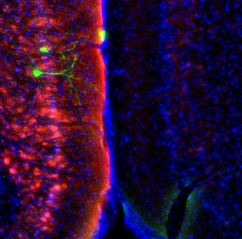
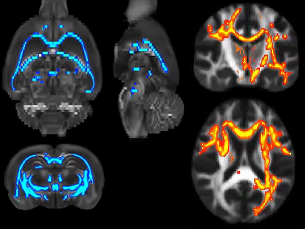
Alcohol dependence is marked by a gradual decline in cognitive control and difficulty adapting to changes. However, the underlying neuroadaptations are still poorly understood. In an animal model of alcohol use disorders (AUD), we investigated the effects of chronic intermittent alcohol exposure on brain microstructure using dw-MRI, immunohistochemistry, and electrophysiological recordings. Our findings revealed that alcohol exposure significantly impacted the microstructural integrity of the fimbria/fornix, leading to reduced myelin basic protein content and impaired communication between the hippocampus (HC) and prefrontal cortex (PFC). Using a quantitative neural network model, we demonstrated how this disrupted HC-PFC communication could hinder the extinction of maladaptive memories, ultimately decreasing cognitive flexibility. To validate our animal model findings, we combined dw-MRI scans and psychometric data from AUD patients. Interestingly, we observed a correlation between the magnitude of microstructural changes in the fimbria/fornix and reduced cognitive flexibility. These findings emphasize the vulnerability of the fimbria/fornix microstructure in AUD and its potential contribution to alcohol-related cognitive impairments. Further research in this area could shed light on alcohol pathophysiology and aid in the development of targeted interventions.
Photo (left to right): Laura Pérez-Cervera, Santiago Canals, Silvia De Santis.


 Español

