Español
Age Impairs Corneal Sensitivity and Reflex Tearing in Congenital Aniridia.
A clinical study led by the Institute for Neurosciences reveals that aniridia causes a progressive loss of corneal sensitivity
• The study, published in the journal Cornea, shows that this rare disease not only alters the structure of the cornea but also affects the function of the sensory nerves that protect and maintain it.
• Through a clinical study conducted in children and adults with aniridia, the researchers found that the loss of sensitivity is not static, but rather progresses with age.
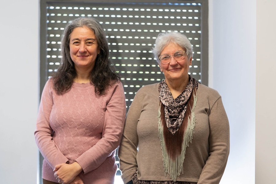
(Photo: Researchers Mª Carmen Acosta and Juana Gallar, who lead the Ocular Neurobiology laboratory at IN UMH-CSIC)
Congenital aniridia is a rare disease caused, in most cases, by mutations in the PAX6 gene, which is essential for the development of ocular structures. Although the most visible feature is the total or partial absence of the iris, its effects go far beyond this, as those affected often experience focusing problems, photophobia, and various complications that may worsen over time. Now, a clinical study led by the Ocular Neurobiology Group at the Institute for Neurosciences (IN), a joint center of Miguel Hernández University of Elche (UMH) and the Spanish National Research Council (CSIC), and published in the journal Cornea, has demonstrated for the first time that this condition not only affects the anatomy of the eye, but also the function of the corneal sensory nerves.
Until now, previous studies have shown that nerve density in the corneas of adult patients with aniridia is reduced. However, no one had analyzed whether these nerves were fully functional: “We knew there were fewer nerves, but we still needed to understand whether the remaining ones were working properly and what consequences this had for the eye”, explains Professor Mª Carmen Acosta, who led the study. To carry out this research, the team evaluated a group of patients with aniridia, including both children and adults, and compared them with individuals without the ocular condition.
Since aniridia is a rare disease, assembling a clinical cohort that included different age groups posed a significant challenge. The study was made possible thanks to collaboration with ophthalmologist Nora Szentmáry, a specialist at Semmelweis University (Hungary), whose research background and clinical expertise in aniridia make her an international reference in this field. Her unit facilitated patient recruitment and evaluation, a key factor in enabling the analysis of how nerve function evolves from childhood to adulthood.
The researchers measured corneal sensitivity to very mild mechanical stimuli applied through controlled air pulses, as well as sensitivity to cold. They also analyzed tear production under basal conditions and after activating the lacrimal reflex through CO₂ microstimulation using the i-Onion device, developed from a patent held by the research group itself.
The results showed a clear pattern: in childhood, corneal sensitivity is very similar to that of healthy individuals. However, in adulthood, a significant decrease appears, and patients require stronger stimuli to perceive contact, showing difficulty distinguishing stimulus intensity. “The most relevant finding is that the deterioration is not static, but progressive. Children still retain a function quite close to normal, but in adults, we observe a clear loss of sensitivity”, Acosta notes.
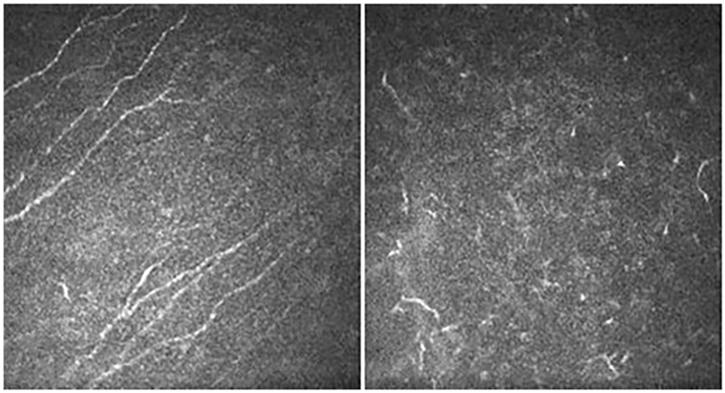
Cornea of a healthy eye (left) and of a patient with aniridia (right), showing changes in the tissue’s defensive cells that reflect corneal alteration. Source: Csorba et al., Current Eye Research, 2024.
The study also reveals alterations in the tear response. Although basal tear production is comparable to that of people without aniridia, the ability to increase secretion in response to a protective stimulus is reduced, limiting one of the eye’s main defense mechanisms. “Sensory information not only allows us to detect contact or cold. It is what activates protective mechanisms such as blinking and tear production. If the nerve signal weakens, the eye’s defense system also weakens”, explains Professor Juana Gallar, head of the Ocular Neurobiology Group.
The cornea loses its ability to regenerate
Another relevant aspect of the study is that the team focused on the trophic function of sensory nerves. Beyond their role in the perception of sensations, these nerves actively contribute to maintaining and regenerating tissue. When innervation decreases or deteriorates, the cornea loses its ability to repair itself, which promotes the appearance of small lesions, loss of transparency, and persistent pain. “Nerves are essential for keeping the cornea healthy. If their function becomes altered over time, the tissue no longer regenerates properly, and complications arise that affect both vision and quality of life”, adds Gallar.
This work is part of a broader project in which the team studies aniridia, both in patients and experimental models. In the next phase, the researchers will further analyze nerve function in a mouse model carrying a mutation in the PAX6 gene, which will allow them to study in greater detail the cellular mechanisms involved in the progressive degeneration of corneal innervation. Understanding these processes at a basic level is essential for designing more precise therapeutic strategies in the future to help slow deterioration and improve patients’ quality of life.
This research was possible thanks to funding from the Spanish State Research Agency – Spanish Ministry of Science, Innovation and Universities, the European Regional Development Fund (ERDF/European Union) “A Way of Making Europe”, and the Generalitat Valenciana. It also received support from the Dr. Rolf M. Schwiete Foundation, the Hungarian Academy of Sciences, and the National Research, Development and Innovation Office of Hungary.
Source: Institute for Neurosciences UMH-CSIC (in.comunicacion@umh.es)